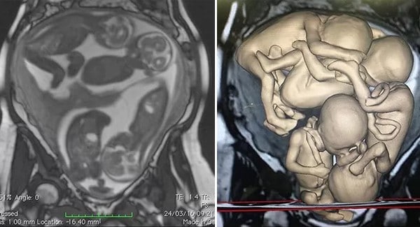
A 32‐year‐old woman with an unremarkable medical history, who failed to conceive after 3 years of trying, was diagnosed with b.ilateral en.dometrioma, peritoneal endometriosis and right tubal dilatation on MRI. The couple were offered assisted reproductive technology (ART). In the first cycle of in‐vitro fertilization (IVF), eight oocytes were collected. This first attempt was unsuccessful and the embryo transfer resulted in a m.iscarriage after 4 weeks. This is called a monochorionic diamniotic quadruplet pregnancy.

A new attempt at IVF was made 6 months after the first cycle. Eight oocytes were obtained and fertilized, resulting in three embryos. Two blastocysts were transferred. Transvaginal ultrasound examination at 6 weeks showed two uterine sacs, each containing two embryos. The couple were informed of the maternal and fetal r.isks of a quadruplet pregnancy and counseled on the possibility of fetal reduction, which was declined.

During a first trimester ultrasound examination, the anatomy of all four fetuses was apparently normal. Cervical cerclage (cervix is closed) was performed at 16 weeks for the prevention of preterm delivery and vaginal progesterone was prescribed at 24 weeks. A fetal MRI was performed at 26 weeks due to difficulty in evaluating complete fetal anatomy with ultrasound, and 3D virtual and physical models were constructed using the data. A c-section was performed at 32 weeks due to uterine contractions and dyspnea, delivering four male babies. Apgar scores at 1 and 5 min were 9 or 10 in all neonates. Two neonates were discharged from the hospital 34 days after delivery and the other two at 36 days after delivery.

Our pr.ayers for those women who are waiting for a miracle in their womb, that G.od grant them the gift of being mothers.


